Recognizing the clinical utility of whole body MR imaging, radiologists at Kawasaki Saiwai Hospital (Kawasaki, Japan) began offering whole body diffusion weighted imaging (DWI) in 2009 for oncology patients. In 2012, the hospital installed a Philips scanner, the Ingenia 1.5T. The dStream digital architecture and highly linear gradients of Ingenia allowed them to switch to coronal – rather than axial – whole body DWI, and were key to developing a fast, high quality protocol that has led to increased referrals and decreased dependence on nuclear medicine imaging.

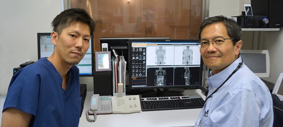
Takanori Naka, MR technologist (left) and Hiroshi Nobusawa, radiologist (right) at Kawasaki Saiwai Hospital, Japan.
High contrast between lesions and background is beneficial in oncology patients
Radiologist Hiroshi Nobusawa, MD, PhD, explains that the coronal DWIBS protocol for whole body DWI is excellent for visualizing lesions in oncology patients. “About 90% of the DWIBS exams are done in this type of patients. The remainder of DWIBS exams are performed to gain information in cases of fevers of unknown origin,” he says. “The DWIBS sequence’s value in oncology cases is due to the high contrast it creates between lesions and surrounding tissue. Whole body DWI is requested by physicians who need to clarify TNM staging or determine therapeutic strategies, oncologists in need of diagnosis or follow-up scans, surgeons who need to see the presence of distant lesions that are sometimes difficult to detect by CT before surgery, and urologists for the evaluation of bone lesions, and the effect of chemotherapy and radiotherapy.”
What is DWIBS?
Diffusion Weighted Imaging with Background Suppression (DWIBS) is an alternative to PET-CT for visualizing lesions throughout the body, supporting the role of MR in oncology studies. DWIBS suppresses normal organ tissue, blood, muscles and fat to achieve high contrast between background and lesions. Moreover, patients can breathe freely during the entire DWIBS study.
Join forces with two industry leaders
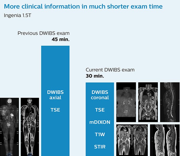
Shorter exam time, better patient acceptance, more clinical information Before the Kawasaki Saiwai Hospital adopted the Philips Ingenia, the long exam length was hindering clinical adoption of whole body DWI. Their first goal after the Ingenia was available was to shorten exam time. The time saved could then be used to fit in more clinical information while keeping the total exam time acceptable for patients. Switching to coronal DWIBS rather than axial further shortened the scan time. Mr. Naka says: “When we use a coronal DWIBS acquisition, we can perform a full whole body examination, including other required sequences, within 30 minutes. This is considerably faster than the previously used exam with axial whole body DWI, which took more than 45 minutes.” This allows the exam to be used on patients in poor health who would have difficulty tolerating a long exam, and simplifies scheduling.

More clinical information in much shorter exam time.
Accelerating acceptance and referrals

The Kawasaki Saiwai Hospital.
With an optimized protocol and the ability to make diagnoses that used to depend on nuclear medicine studies, acceptance in the Kawasaki Saiwai Hospital has increased. And with active education of referring physicians, referrals are also on the rise. Dr. Nobusawa says, “As soon as you understand the usefulness of DWIBS exams with the Ingenia system, surely you would like to use it. We hope that DWIBS once will be adopted as a gold standard in the care for oncology patients.”
Clinical case: Whole body MRI of bone lesions in spine
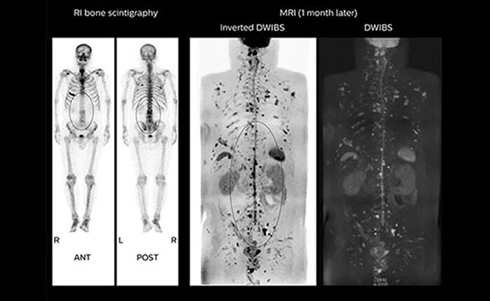
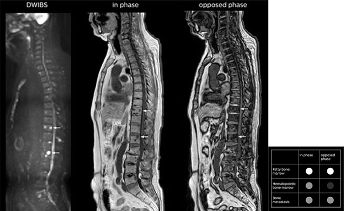
This oncology patient received diagnosis extent of disease (EOD) grade 2 after bone scintigraphy. The lesions in the spine (RI bone scintigraphy) were thought to be age-related bone marrow effects. When the patient was sent to MRI one month later, he was scanned with the whole body oncology protocol. Many bright bone lesions were seen (inverted DWIBS) on the DWIBS images. The Dual FFE in-phase and opposed-phase images can help the physician to further characterize the lesions. Fatty bone marrow would be expected to appear bright on both in-phase and opposed-phase images. Hematopoietic bone marrow would be expected to appear mid-gray on in-phase and dark on opposed-phase images. The lesions in this patient are mid-gray on both in-phase and opposed-phase images. The therapy plan for this patient was changed from radiotherapy to pain relief in line with EOD grade 4.


For more information
This customer story was excerpted from a longer case study in FieldStrength magazine. Read the full article, including more clinical cases, here

